


Ajay K, Venket Shelke and Partha Das, Kemin Industries South Asia Pvt. Ltd.
INTRODUCTION
Bones play a vital role in supporting muscle mass, protecting vital organs, and regulating calcium and phosphorus content in the blood. With the advent of intense genetic selection to achieve rapid weight gain, lameness or leg weakness became a burning issue in today’s poultry industry across the globe. Lameness negatively affects both animal welfare and farm profitability. Lameness is the major reason behind reduced growth, increased culling rate, mortality, poor uniformity, and condemnation of chicken meat. The lameness condition is multifactorial. Although the rapid growth rate is considered the main factor for lameness, factors like management practice, disease, nutrition, genetics, toxins, sex, and age are also responsible for the condition. Thus, it becomes essential to differentiate between these factors to develop proper prevention and treatment strategy. Classification of leg disorders is hard, as they are multifactorial and sometimes factors are interconnected. However, leg disorders can be broadly due to infectious and non-infectious causes.
A. INFECTIOUS CAUSES
♦ Bacterial chondronecrosis with osteomyelitis (BCO)/ Femoral head necrosis (FHN)/ Bacterial chondronecrosis (BCN)/ Tibial head necrosis (THN)
BCO can affect the proximal femur, tibiae, and flexible thoracic vertebrae. This is mostly seen in 25 to 45 days old broilers. In fast-growing birds, mechanical stress on the growth plate produces microfracture which is colonized by opportunistic bacteria via translocation through the gastrointestinal tract (GIT), respiratory tract, or skin and results in BCO. The BCO can be broadly classified into vertebral BCO, FHN, and THN.
♦ Vertebral BCO presents nodular swelling and abscessation of the 4th thoracic vertebral (T4) body with osteomyelitis and compression of the spinal cord. Lung tissue is focally adherent to the abscessed vertebrae.
♦ FHN–Necrosis of the femur head due to colonization of opportunistic bacteria results in the separation of the growth plate of the proximal femur from its articular cartilage.
♦ THN–When the necrosis area comprises the proximal tibial part, it is called tibial head necrosis.
Clinical signs: Hock-sitting posture and the reluctance to walk. When birds walk, they place their wing tips on the ground and limp gait in the advanced stage. Birds sound loud when the proximal femur was palpated.
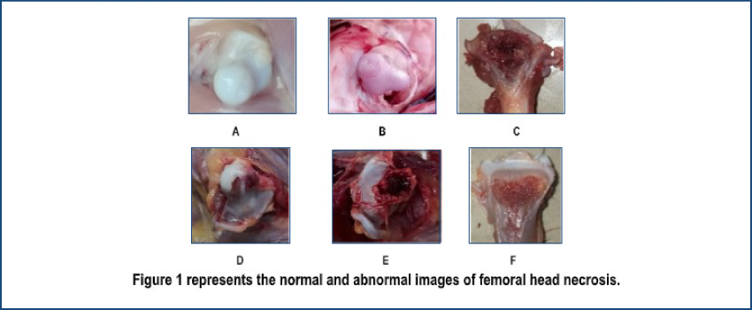
Note: A–Normal Femur head; B–Mild FHN; C to E–Severe FHN; F–Mild THN
Lesions: Vary from small pale areas adjacent to the growth plate to large zones of yellow tissue extending from the growth plate to the medullary cavity.
Etiology: Mainly by Staphylococcus aureus, Enterococcus cecorum, and Escherichia coli. Immunosuppression by Reo, infectious bursal disease (IBD), chicken anemia virus, adeno, and pox virus is highly associated with an increased level of BCO.
Prevention: Feed-grade probiotics starting from day-old chicks, antibiotics like enrofloxacin and chlortetracycline, hatchery and farm hygiene, water hygiene, and maintaining immune status to the optimum level.
♦ Tenosynovitis and Arthritis
Tenosynovitis is an inflammation of the tendon sheath. This can happen by bacteria, mycoplasma, and viral infection. Reovirus, Mycoplasma synoviae, Staphylococcus aureus, and retroviruses are pathogens responsible for this condition.
Clinical signs: Mild to moderate lameness, hot swollen tendons, and joints. Affected joints when cut open present vicious grey to translucent exudates in mycoplasmal infection unlike opaque exudates in other bacterial infections.
Prevention: Chemotherapeutic agents like anti-mycoplasma drugs in farm and breeder flock, dipping of hatching eggs in an antibiotic solution, vaccine at breeder flock, and water hygiene.
♦ Infectious stunting syndrome (ISS)/ Helicopter disease/ Brittle bone disease/ Pale bird syndrome/ Malabsorption syndrome/ Runting and stunting syndrome
Mainly reported in young chicks.
Clinical signs: Stunted growth and swollen joints, stunted chicken which remains small despite voracious appetites. It becomes more noticeable after two weeks when the unaffected chickens grow rapidly. Impaired digestion resulting in poor absorption of nutrients is one of the reasons because of enteritis evidence.
Etiology: Reovirus
Prevention: Probiotics, high doses of vitamins A, D, and E, cold stress management, vaccination at breeder flock, antibiotics to control secondary bacterial infection, hatchery, farm, and water hygiene.
♦ Avian Encephalomyelitis
Avian encephalomyelitis (AE) is a disease in chicks up to two to three weeks of age. AE is a viral disease affecting the central nervous system of young chickens. Signs include tremors, ataxia, leg weakness, and paralysis of both legs extended out to one side. Lesions sometimes include gray to white foci which may be visible on cut surfaces of the muscle in the gizzard. AE must be differentiated from encephalomalacia, which can cause ataxia that might be due to the deficiency of vitamin E.
Prevention: Vaccination of breeder flock.
♦ Osteoporosis/ Cage layer fatigue
This condition mainly occurs in laying hens in peak production due to decreased mineralization of long bones. Signs include paralyzed birds laying on their back in cages. During postmortem, one may find the shelled egg in the shell gland and fragile or brittle bones. Decreased mineralization may be due to calcium, phosphorus, and vitamin D deficiency.
Prevention: Phase-wise feeding with ideal calcium level.
♦ Amyloidosis
Amyloidosis mainly occurs in adult layer birds. It is associated with chronic infectious bronchitis (IB) infection or administration of repeated multiple killed vaccination. Affected birds are characterized by the presence of amyloid protein in joints and internal organs like the liver and spleen. The affected joints are swollen and contain orange-yellowish material. Signs include muscular shivering and lameness. Post mortem lesion includes edema of pectoral muscles, congested ovules in layer birds, and other classical lesions as found in IB.
Prevention: Live vaccine of appropriate sero type of IB and mitigating chronic inflammation.
♦ Marek’s Disease
Marek’s disease is caused by the herpes virus which causes paralysis and immunosuppression. Swollen peripheral nerves like sciatic nerves result in coordination and progressive lameness. Affected birds show typical posture with one leg starched forward and the other backward.
Prevention: Vaccination of breeder flock.
B. NON-INFECTIOUS CAUSES
These disorders include developmental, degenerative, and metabolic disorders. Genetic selection, growth rate, feed conversion efficiency, exercise, stocking density, nutrition, management, sex, age, and growth rate are important factors responsible for these disorders.
♦ Varus valgus deformation (VVD):
The most common long-bone distortion is seen in commercial broilers with normal ossification. Valgus deformity produces bow-legged and varus produces knock-kneed stance. Distal tibiotarsus is the common site of angulation. These conditions may be with ligament and tendon rupture or displacement.
Clinical signs: Moderately affected chickens walk with a waddling or hobbling gait, while severely affected birds are either unable to walk or walk on their hocks.
Etiology: Biomechanical forces. Genetic defects, high growth rate, weight-bearing, lack of exercise, and hard floor.
Lesions: No thickening of the growth plate and angulation at the distal tibiotarsal part.
Prevention: By genetic selection, slowing the growth rate by early feed restriction, and shortened photoperiod.
♦ Rotated tibia
In broilers with the rotated tibia, the tibiotarsus is often rotated through the long axis and generally externally through 90 degrees, so that the lower leg is abducted, and the bird is described as having spraddled legs. Incidences are very less. In this case, the tendon is not displaced and deformity is unilateral.
Clinical signs: Spraddled legs.
Etiology: Biomechanical force, biotin deficiency, genetic selection, and stocking density.
Prevention: Feed restriction and a step-up lighting program reduce the incidence of the rotated tibia.
♦ Tibial dyschondroplasia (TD)
Insufficient angiogenesis, failure of chondrocyte hypertrophy, and delay in cartilage calcification are major factors that lead to TD.
Clinical signs: Swelling and bowing in the region of knee joints, angulations of legs, typically in birds >35 days.
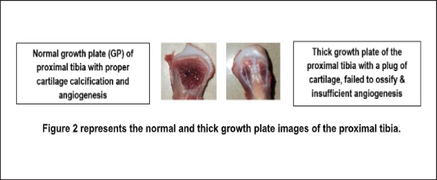
Etiology: Calcium (Ca)/ Phosphorus(P) ratio, excess plasma chloride, copper deficiency in feed, metabolic acidosis, mycotoxins like fusarium, thiram toxicity, salinomycin toxicity, hydrogenated soybean oil, and genetics.
Lesion: Thickening of the growth plate of the proximal tibia and plug of cartilage (non-mineralized cartilage) in the proximal end of the tibia.
Prevention: Correction of imbalance diet, broad spectrum toxin binder, taurine to correct tibial dyschondroplasia induced by thiram toxicity, correcting dose of salinomycin.
♦ Spondylolisthesis
The clinical manifestation of T4 subluxation is known technically as spondylolisthesis (spine slippage) or as “kinky back,” in which spinal cord compression leads to paraplegia, a hock or tail sitting posture, and an inability to stand. This condition is non-infectious in origin. It occurs in 2-4 weeks of age as flexible thoracic vertebrae are not fully fused.
♦ Pododermatitis/ Contact Dermatitis/ Hock Burns/ Ammonia Burn
Contact dermatitis is a disorder on the skin of foot pads, hocks, and breasts. Foot-pad dermatitis is also known as bumblefoot. Birds are seen with necrotic lesions on the plantar surface of footpads. Birds with severe foot pad lesions are lame.
Etiology: Wet litter due to water spillage near drinkers, high stocking density, diarrhea, loose droppings, high ammonia, humidity, and high dietary protein.
Prevention: Good litter management, improving ventilation, and biotin supplementation.
♦ Ionophore Toxicity
Ionophores like monensin, salinomycin, and lasalocid may induce lameness if used in excess or with tiamulin combinations. Monensin and salinomycin induce paralysis with both legs extended backward, while lasalocid induces lameness by walking in a toe-like pattern. Severe myodegeneration occurs in these types of toxicity.
Prevention: Avoid overdosing on ionophores and mixing errors.
♦ Metabolic Disorder
⇒ Rickets: Soft bones occurring in young birds due to reduced mineralization is known as rickets. Rickets can occur in both fast-and slow-growing chicks. Rickets can be hypocalcemia rickets or hypophosphatemia rickets.
Clinical signs: The birds are alert and bright in appearance but often recumbent and remain sitting on their hocks with extremely pliable beaks and long bones. Bones are easily breakable. It is manifested by enlargement of the end of the tibia (rachitic lesions in the tibiotarsus) and femur with wide and irregular growth plate and beading of ribs at the joint.
Etiology: This disorder is usually caused by abnormal Ca/P ratios in the ration, low Vitamin D3 levels in the ration, excess dietary magnesium and iron, mycotoxicity, or intestinal malabsorption associated with enteric diseases.
Prevention: Feed with balanced calcium and phosphorus with Vitamin D3.
⇒ Chondrodystrophy/ Perosis/ Slipped Tendon: Chondrodystrophy is a disorder of the growth plates of long bones, where longitudinal growth is impaired while mineralization and appositional growth remain normal. Generally occurs in young birds. Nowadays, these conditions are not seen frequently due to a well-balanced diet but outbreaks may occur due to feeding supplement and feed additives mixing errors.
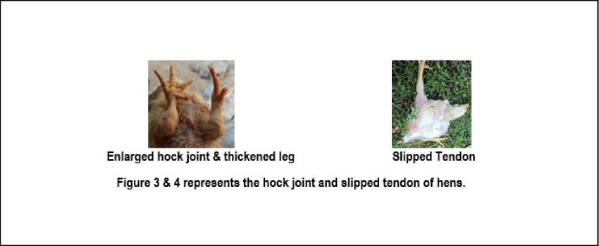
Clinical signs: Thickened and shortened legs, enlarged hock joint with normal mineralization and thick growth plate without a plug of cartilage, bending of the distal end of tibia, in severe cases, the tendon may get slipped.
Etiology: Trace mineral deficiency of manganese (Mn), zinc (Zn), calcium (Ca), and phosphorus (P) imbalance, Vitamin D3 and E, choline, folic acid, pyridoxine, niacin deficiency, and a high protein diet deficient in other vital nutrients.
Prevention: Mn through water or feed, Zn, Ca, and P balance, ensuring proper feed additive mixing.
⇒ Curled Toe Paralysis: This condition arises due to a deficiency of riboflavin (Vitamin B2) in the early days of a chick. Due to damage in peripheral nerves, affected chicks sit on their hock with toes curling inwards and downwards.
Prevention and Treatment: Water soluble vitamins containing Riboflavin and Vitamin C can help in nerve regeneration. The dose of vitamin C for chickens is 250 mg/kg body weight.
CONCLUSION
Genetics, management, and growth rate are important factors for preventing leg weakness and lameness-related issues in modern broilers. Bacterial Chondronecrosis with Osteomyelitis, Tibial Dyschondroplasia, Pododermatitis, and Varus Valgus Deformation are the most common disorders in today’s modern broiler farming. Improving gut integrity and immunity by feeding CLOSTAT™, the next-generation probiotics, and Aleta™, an algal-based immunomodulator may help mitigate the severe issue of BCO and other conditions like dermatitis, bacterial arthritis, and pododermatitis. A balanced diet plays a crucial role in preventing Tibial Dyschondroplasia, Chondrodystrophy, and Rickets.